RSA Introduction
Radiostereometric Analysis (RSA) is a high precision method for
measuring micromotions in the skeleton (Selvik
74,89, Kärrholm 89).
Clinical use of RSA may be divided into the following steps:
- Small spherical markers (0.5-1.0 mm in diameter) made of
Tantalum are inserted into the patient and/or attached to the
prosthesis. The markers are grouped into segments, which
may be used to define an orthopaedic entity, e.g. the
femur. Insertion may be done during surgery, but also
non-surgically with an insertion device.
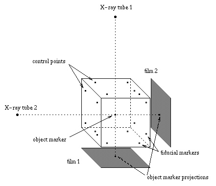
- The patient is X-rayed from two angles simultaneously together
with a reference cage, producing two radiographs. See the
illustration below.
- The position of the marker projections are measured on the films.
- The projected positions of the fiducial markers and
control points attached to the cage are used to
establish the exposure geometry, i.e. the relationship between
the X-ray foci, the reference cage, and the X-ray films.
- The three-dimensional coordinates of the patient markers are
reconstructed from their projections.
- Given the result of a previous RSA examination, the motion
between different segments may be calculated.
Last modified: Wed Mar 4 15:30:59 MET 1998